Translate into Your Language
Fibrelos Rock Clots
Fibrelos Rock Clots: See all the Diagrams
Fibrelos Rock Clots assist the Fibrelos Tangles to build in size. When you see the Little Black/Bluish/Gray  Dots on the upper epidermis, that's the Fibrelos Rock Clots. It's clear to see in the photos on this page, as well as in other photos on our website, the Fibrelos Rock Clots are definitely there, and cause quite a problem in the body.
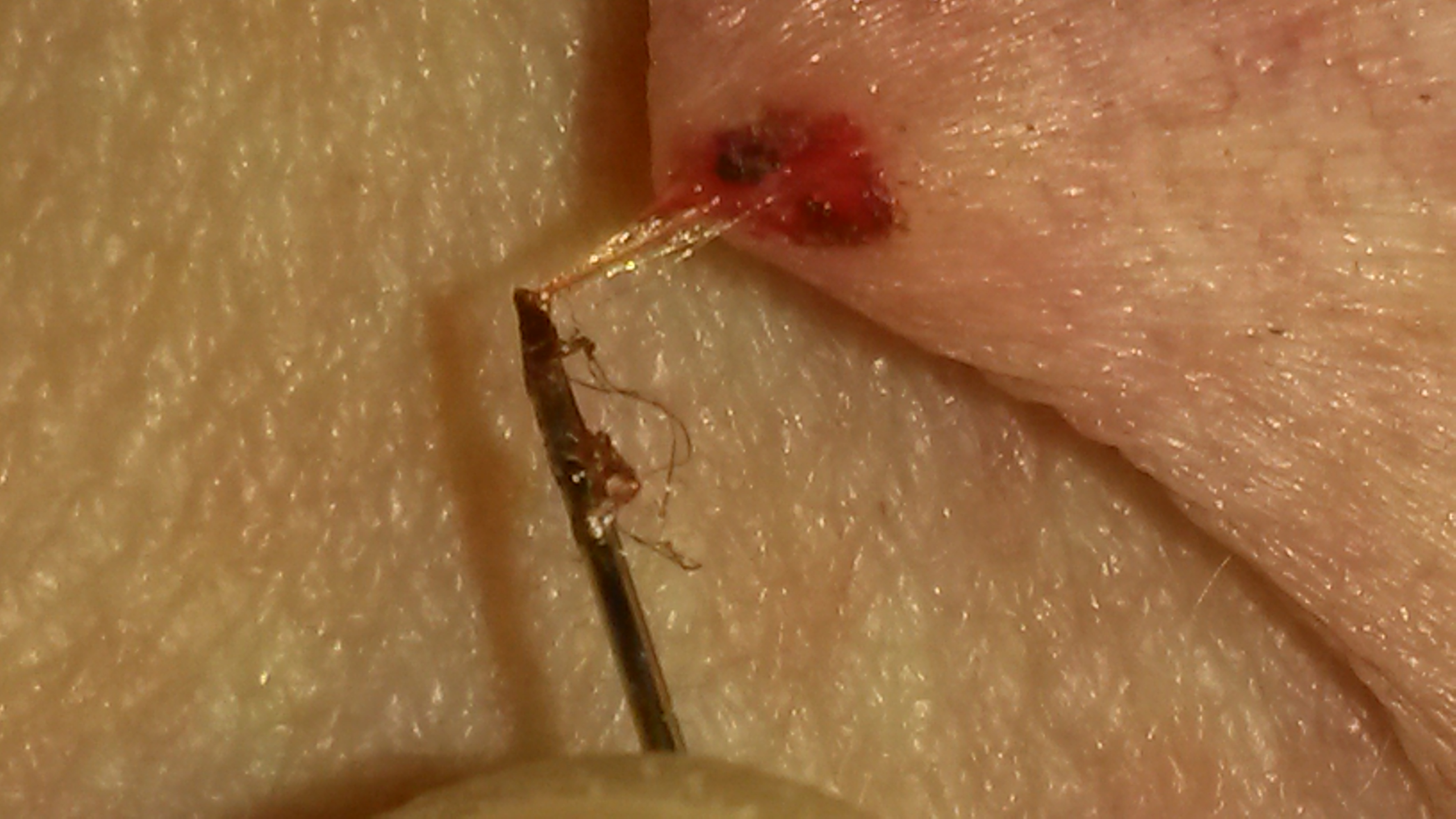
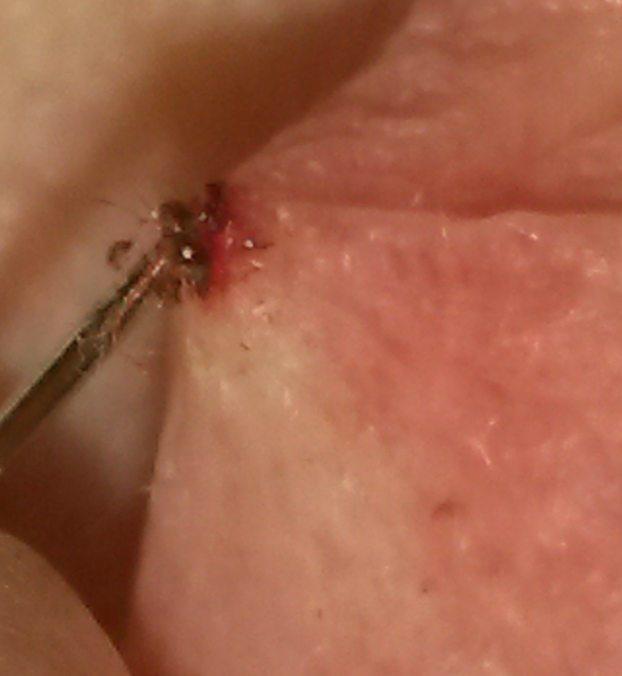
In the diagram to the right, you see a Fibrelos Rock Clot being untangled from the Fibrelos Strands. The location at the chest toward the heart approximately 2" to the right of the heart. In the diagram below, it shows the Pigmentation location with the raised area in the other diagrammed photo directly below . There were some heart palpitations getting stronger, and once the raised area plus the Pigment on top was seen, our Founder knew exactly what was happening. This is problematic due to the palpitations felt by the person, yet not showing up this activity on an EKG.
Through the top layers of skin, majority of the time, it's visible to see the Fibrelos Rock Clots; however, sometimes they're there, yet could be hidden in a Fibrelos Bump that's the same color as the skin; looking like just a simple raised bump. There's many times, the Fibrelos Rock Clots will be mixed in a mass of Fibrelos Strands tangled underneath the surface; then may show slightly; like a shadow when we look at our skin, there may be a gray color or even look similar to a liver spot, or a different color tone. It's difficult at times to determine, unless you've worked with Fibrelos skin issues, as to the determination of what's going on, this information will be found in this Fibrelos Study. In the diagram above, in the center of the orange square, it shows a bit more about the location where entry of the ©Pin Prick Shunt was made. The other photos placed here, yet right now, showing what came out once entry was made at the middle of the chest, 2" above the heart, and slightly to the right, was a Fibrelos Rock Clot Mini-Tumor. It takes experience to understand exactly what content will be under the epidermis, that's why sharing this with the Public is important; doctors must realize this is a very important piece of medical history being shown & explained.
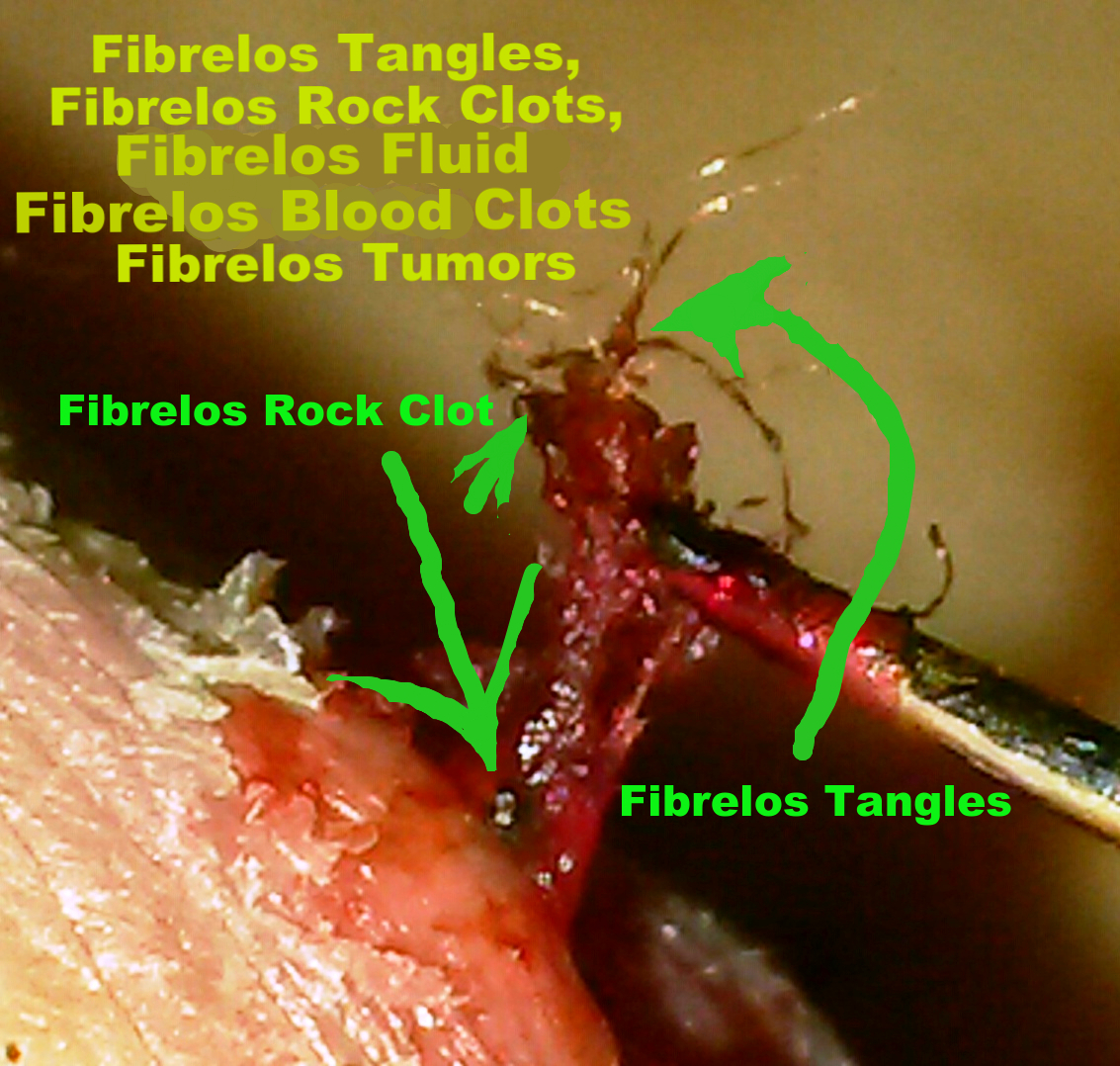
As mentioned above, there's sometimes 'Pigmentation's' showing on the upper layers of the Epidermis that usually are the size of a piece of pepper or even as large as a tomato seed. This showing of Pigment, as with this Fibrelos Study, is cause for concern with what's underneath the surface. Each time we've entered into these areas, there's usually a large amount of Fibrelos Rock Clots tangled with Fibrelos Strands with Fibrelos Fluid. Many times, but not every time, Fibrelos Blood Clots will flow out. If no Fibrelos Blood Clots come out, more than likely, as we've found with experience, the flow of Fibrelos Blood Clots will possibly be located in a different area. Yet it could also mean if pressure or pain of some type is felt by the Patient, more work may have to be done to remove the Fibrelos Strands and any  Fibrelos Tangles in that specific area until the pain or pressure goes away. Next, given that more removal in that specific area may need to be done, the Fibrelos Blood Clots could also be there, depth may need to be 1/2mm or so further with a ©Pin Prick Shunt, then the Fibrelos Blood Clots could flow. It depends on what the Patient is experiencing. Keep in mind that most Radiologists and Doctors will have no idea what this looks like under any Radiological studies, such as an MRI or Ultra Sound. Also, keep in mind that pain & pressure felt by the Patient won't show in an EKG, as proven with our Founder's experience, so if their discomfort is felt, this ©Pin Prick Shunt procedure may possibly be the only way to relieve the pain & pressure. Keeping in mind that the clogging of any shunt could be possible, as it was the clogging of shunt with Fibrelos Rock Clots & Fibrelos Strands that sometimes wrap around; stopping the flow of a shunt. At times, the small Fibrelos Rock Clots may stop a shunt from working proper. When shunt flow is moving, a reduction of pain will prove in time. Research remains to be seen until we have Research Specialties involved, we will only be able to put the information we find to make it available for diagnosing possibilities. We need to build our Research Team. Send us an email if you would like to join our Research Team - Researchers contact us: fibrelosstudy@gmail.com
The Fibrelos Rock Clots are comprised of a combination of Fibrelos Fluid, Fibrelos Tangles, trapped blood, and are sometimes large enough to call 'Miniature Fibrelos Rock Clot Tumors'. They form directly on the Fibrelos Strands, plus flow out loose during removal of Fibrelos Strands, Fibrelos Tangles, Fibrelos Blood Clots and Fibrelos Fluid. Once entry is made with a ©Pin Prick Shunt, it's very common to see Fibrelos Rock Clots in the layers of 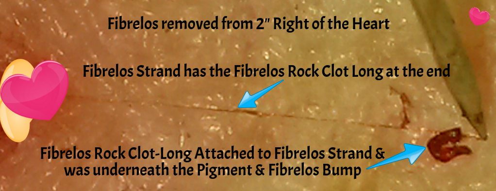 localized tissue under 4x magnification. Keep in mind, we only enter in through the epidermis with a 1/2mm to 2mm depth; there's no need to do anything further, usually, and the virtue of patience is needed with any Fibrelos removal. It takes sometimes two (2) to eight (8) hours for an extensive area to be finished, and even then, you must give the area time to heal; as we have experienced, a twenty-four (24) hour period is needed to allow the remaining Fibrelos Products to maneuver in place of what you've just removed. Sometimes you may also need to allow a few days for proper healing. It's then where the Patient will notice either a full relief, or indicate there's still pain & pressure in the area just worked on with the ©Pin Prick Shunt.
Fibrelos Strand with Fibrelos Mini Rock Clot Tumor at Each End, Between the Breasts
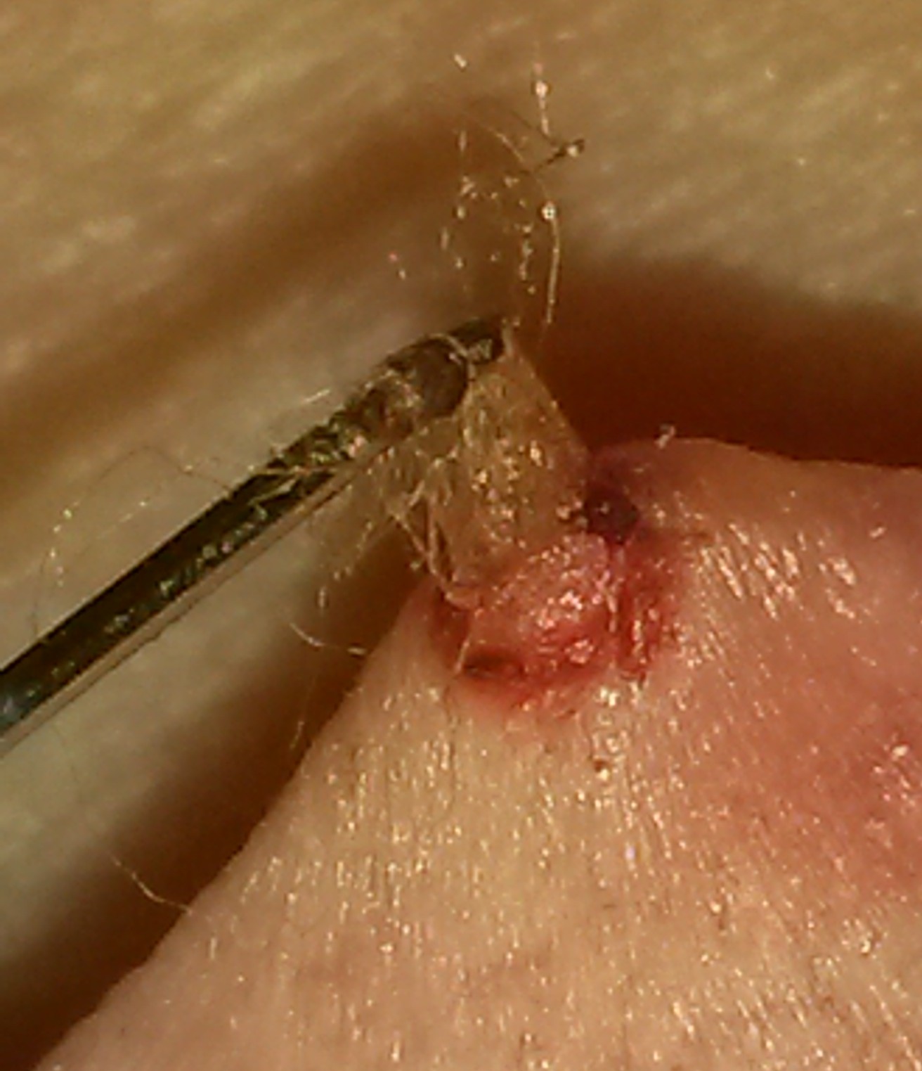
This specific Fibrelos Strand below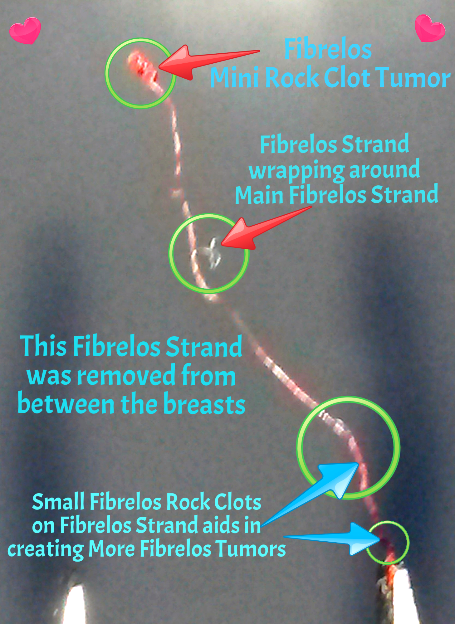 , was removed from between the breasts at cleavage, where heart palpitations were felt very strong. The location of this was to the left of the center of the breasts; and clear to see it was a bluish color Pigment & Fibrelos Bump. After the removal; as more Fibrelos Strands & Fibrelos Rock Clots were removed, the palpitations and pressure significantly decreased.
Note: This is the same Fibrelos Strand as seen below in the other diagram to the left, the photo below was taken a few hours later.
.jpg?timestamp=1541991933808)
Diagrams:
Post Gallbladder Surgery, 8-10-2015
Fibrelos Tangle Bundle: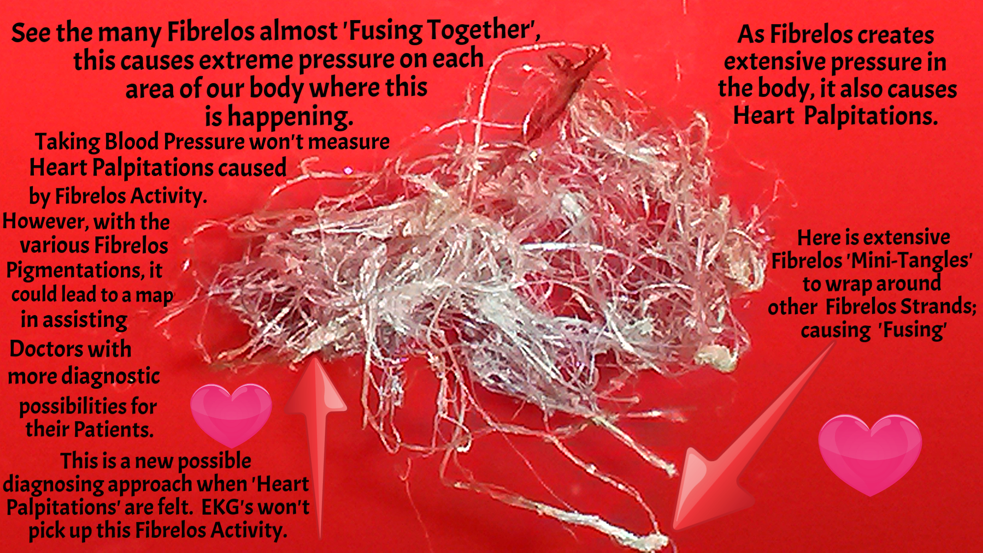
This Fibrelos Tangle Bundle was sent to our first Research Team in October of 2017. As you can clearly see, something like this forming in the body will create problems; as the Fibrelos Tangles form, they also strangle different parts of the body. You can see the 'Fusing of the Fibrelos Strands' together, as the diagram is explaining different facets of each detail, please take a further look at our website in sections to learn more about the devastation of our Founder's suffering. Seen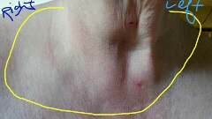 in the strangling of our Founder, tap here or on the photo to take you to the section showing our Founder getting incredibly worse by the Fibrelos in her neck. See the Right side, where pockets of skin travel around the neck; this and the other areas of her neck became worrisome with swallowing, breathing was getting difficult, pain of talking was getting worse. The quick thinking of our Founder led to the relief of a stroke, plus she received relief of the symptoms of swallowing problems with the ©Pin Prick Shunt. Tap the photo and it will take you there.
Center Neck Fibrelos Strands Removed November 3, 2018~Pressure Build Up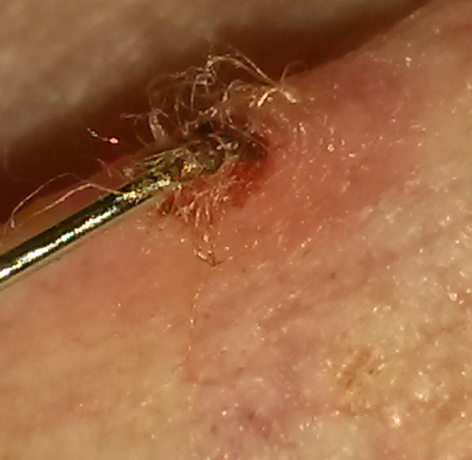

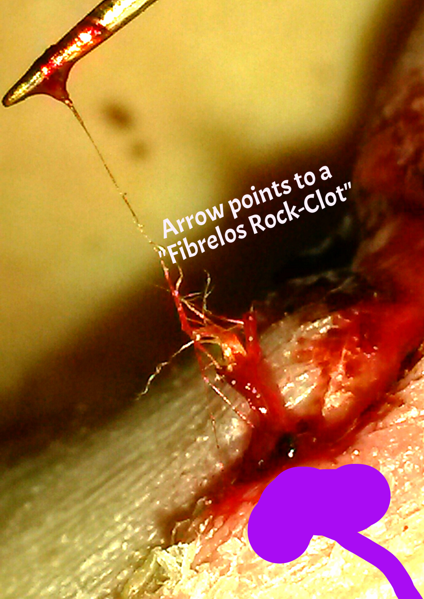
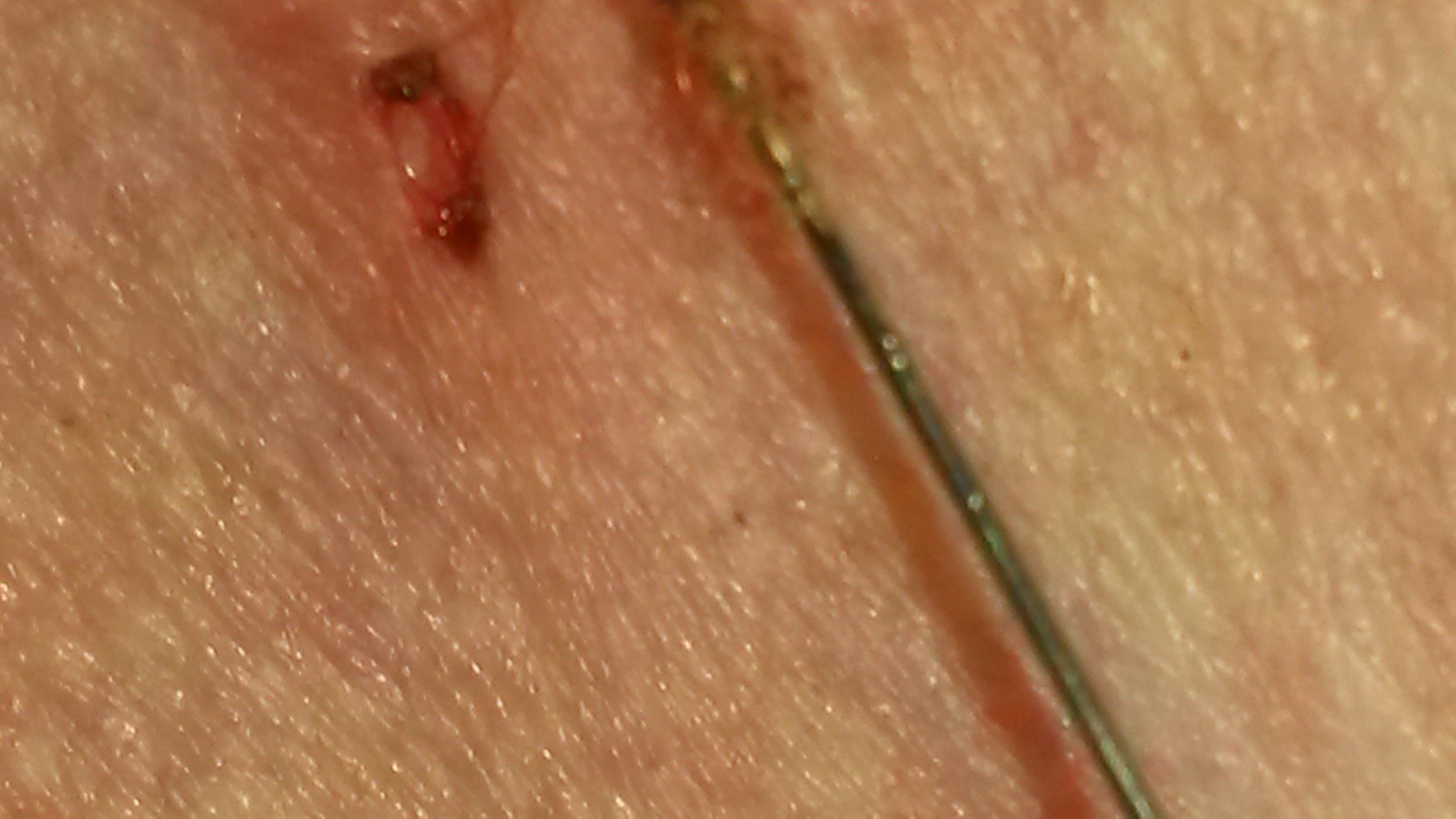
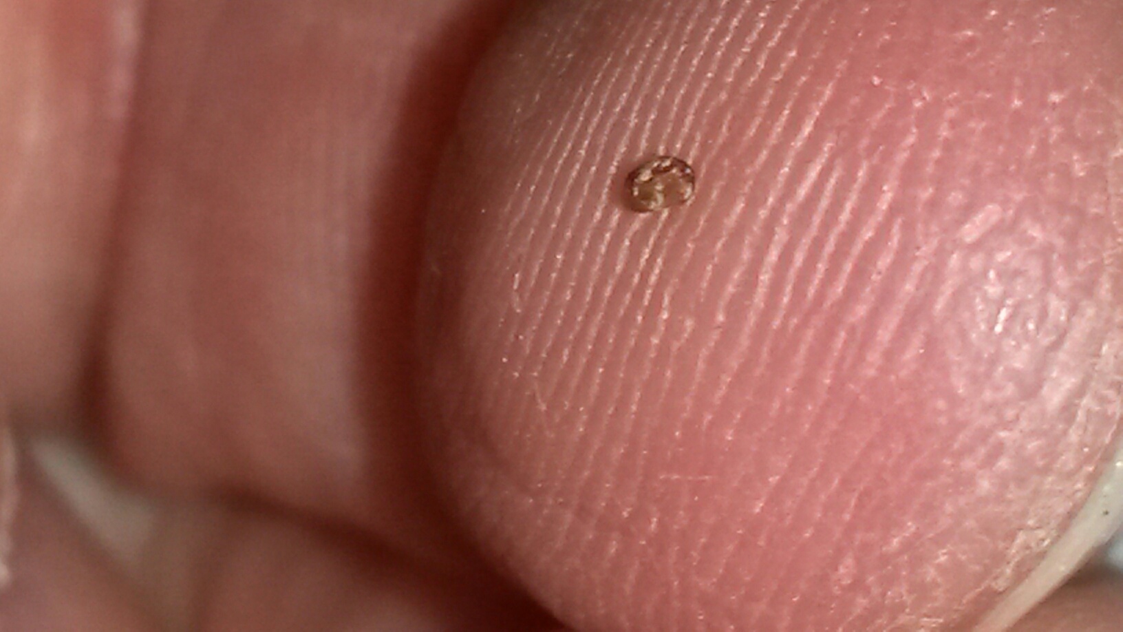
Not A Tick, But Looks Like It...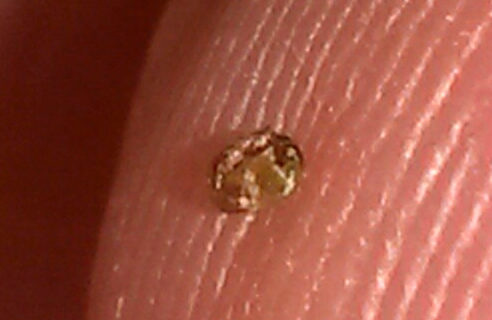
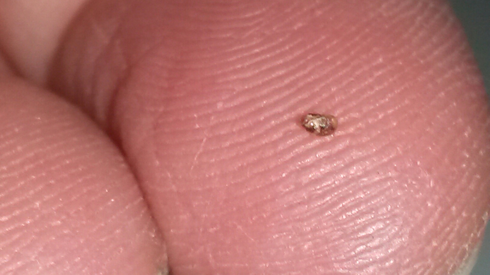
Below you see some similarities in a possible Tick, yet they aren't Ticks at all...these are Fibrelos Mini Tumors. The diagram below also shows a Fibrelos Rock Clot up close; it could also look like a Tick underneath the top layers of skin, yet it's a Fibrelos Mini- Tumor.
 White Flower Productions Copyright for this site: 2000 - 2022
All media is protected under this ©Copyright
©2000 - 2022 Copyright "©The Branchworm Study - Phase I" , "©The Fibrelos Study - Phase II" & "©The Fibrelos Dry Study", "©Fibrelos" is under Copyright ©2000 - 2022 for the ©Fibromyalgia Lyme Disease Foundation (©F.L.D.F.), "©The Pin Prick Shunt" ©2000 - 2022 is under Copyright for the F.L.D.F. ", ©The Natural Flowing Biopsy", ©2000 - 2022 is under Copyright for the F.L.D.F. , ©2018 -2022 The Lyme-No-Mo & Lyme No More Campaigns are Presented by the F.L.D.F. 2020-2022, Copyright Fibrelos Airborne Particle Research Abstract ©2020-2022