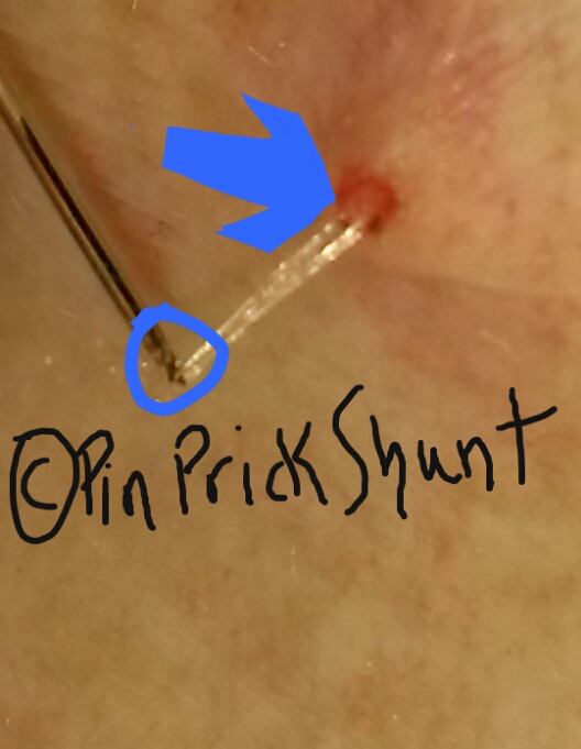
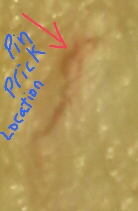
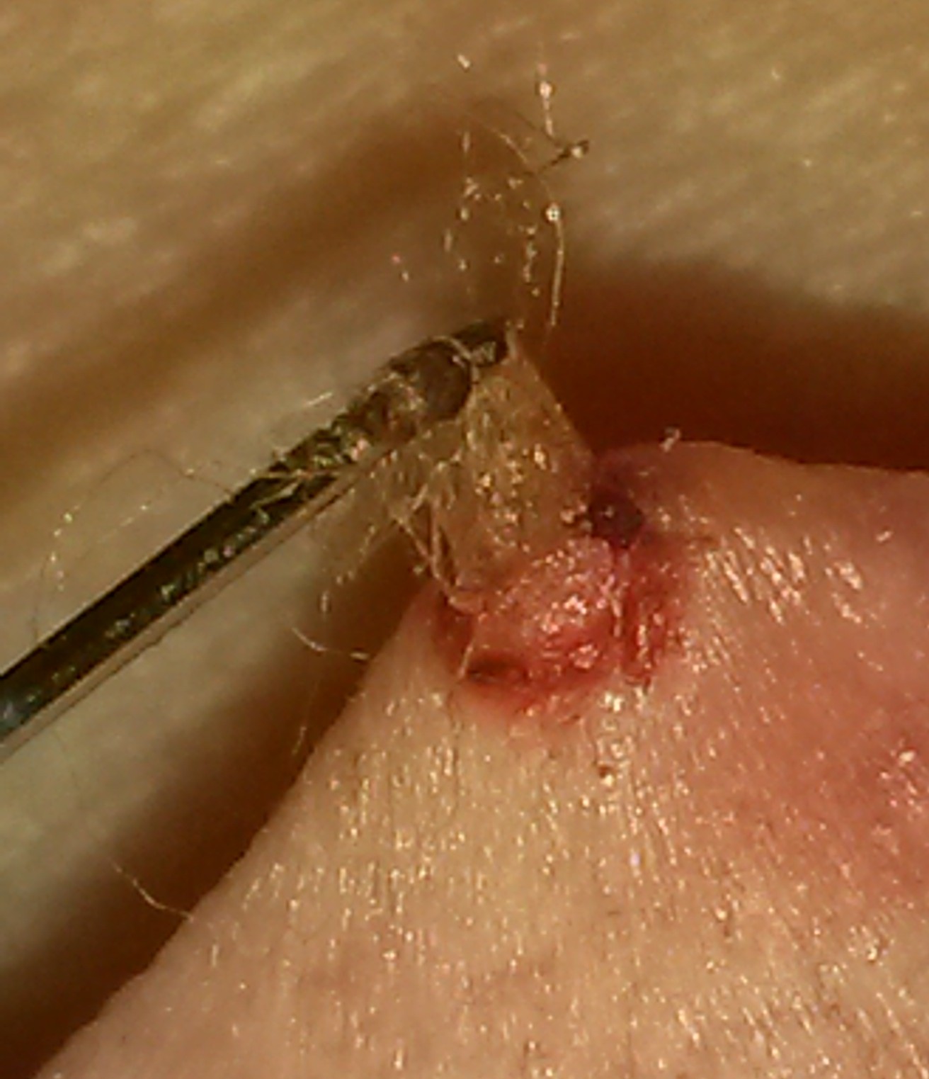
Note: The Blue Circle is the Pin Prick Shunt Tool. The Blue Arrow is pointing to the Shunt Opening just made..jpg?timestamp=1554100203963)
<---The ©Pin Prick Shunt, simply putting it, the way we begin to remove Fibrelos Strands. When a Fibrelos Bump is noticed, this type of ©Free Form Biopsy allows more precision of getting the contents of Fibrelos Strands and other
Fibrelos Products released from the Fibrelos Bump----> 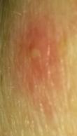
<-----Fibrelos Pressure Bruise 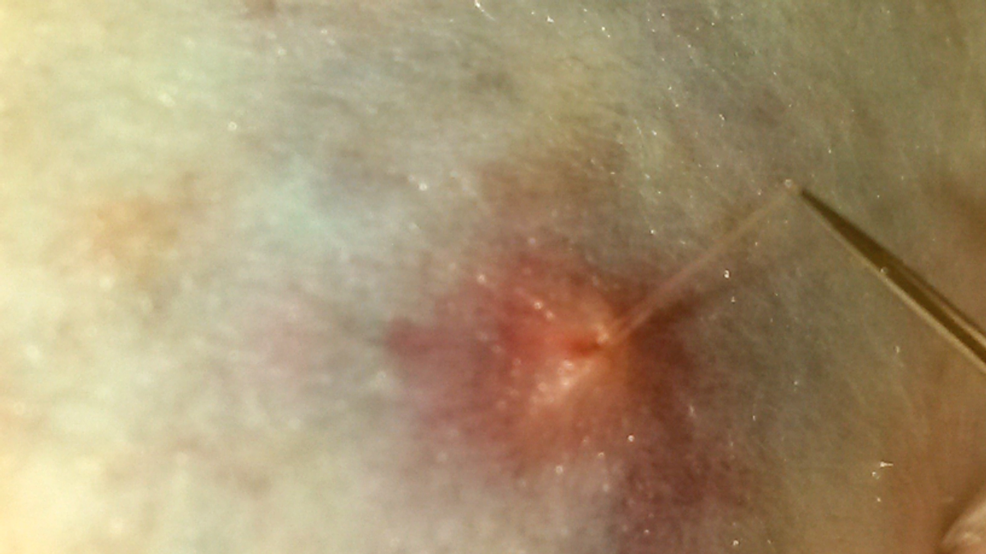 with the assistance of the ©Pin Prick Shunt, Fibrelos Strands are being untangled by using this shunt style. See more below.
The ©Pin Prick Shunt
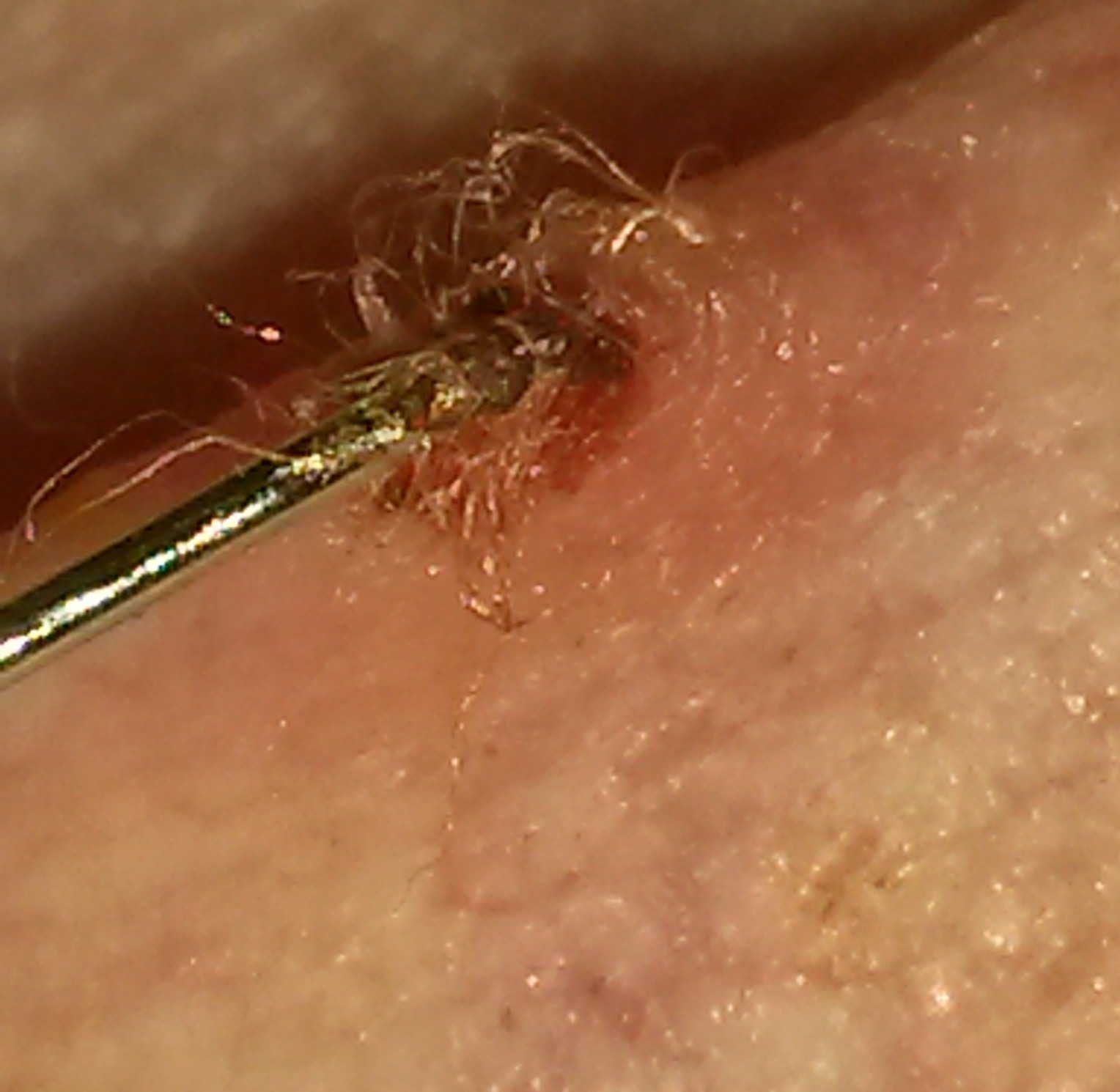
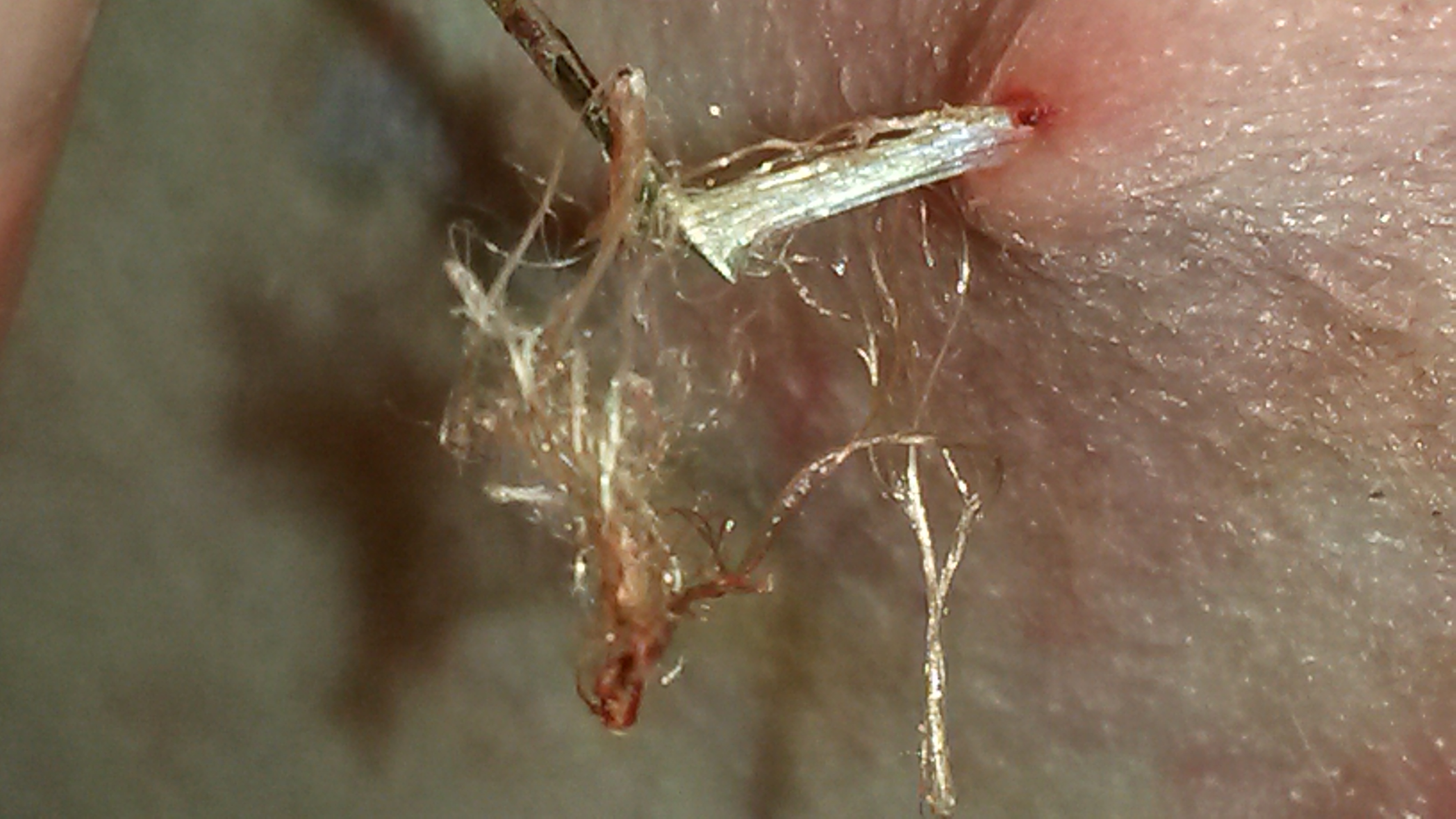
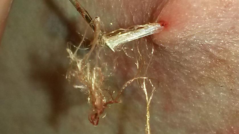 The ©Pin Prick Shunt is a device and technique designed exclusively for the Fibrelos Study (Therer's a video from 3-31-19 where the Pin Prick Shunt relieved extensive chest pressure a little above the heart. Palpitations were being felt by the Founder. We developed a way to biopsy samples with our ©Free Flow Biopsy Method, the sample comes out in a more natural form. This allows each portion to flow out naturally either w ith the Fibrelos Fluid, through blood and/or tangled among Fibrelos Blood Clots and flow. In the photo to the left, this is a mass of Fibrelos Tangles released & removed by using the ©Pin Prick Shunt.
The release of pressure is usually done with an approximately 1/32mm to less than a 1mm tap of the ©Pin Prick Shunt through the skins' surface, but no further than 1mm. This allows a natural flow of release normally not used in medicine today. With this technique immediate relief could be within seconds. In the relief felt by the release of pressure, there's a possibility the pressure could have been building there for an extensive period.
Our website shows firsthand the relief of a ©Pin Prick Shunt. As extensive buildup of Fibrelos Fluid, Fibrelos Blood Clots, Fibrelos Strands, Fibrelos Rock Clots 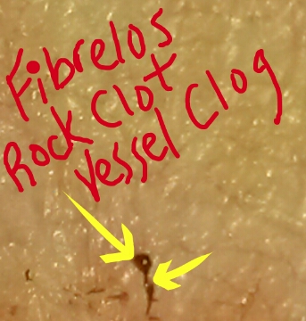 caused by Fibrelos symptoms, the ©Pin Prick Shunt remarkably has shown to be efficient relief.
Because Fibrelos Strands grow in different directions, tangling around forming either a small blockage, a Mini-Fibrelos Tumor that builds with combination of Fibrelos Rock Clots tangling with Fibrelos Strands, and/or a larger tomor possibly to the size of the Fibrelos Basketball Tumor. The tangling of Fibrelos could lead to Fibrelos Mini-Tumors. Usually starting from Tangles of Fibrelos, a mixture of Fibrelos Fluid forming more solid texture to a hardening, breaks up in 'Pepper Size' and/or smaller pieces. The forms of different shapes circular, oval, usually rounded but sometimes flat & sliver like. This is what a Fibrelos Rock Clots is, then it begins growing & forming into a Fibrelos Tumor, Fibrelos Blood Clot forming in different parts of the body. Many times causing a superficial type of thrombophlebitis look of bruising we call this a Fibrelos Pressure Bruise. The Fibrelos Pressure Bruise has been found in the breast tissue, upper leg, and even found on the arms. The Founder has had many of these, and one specific one that centered around a large Fibrelos Bump where a mammogram picked up the bright look of Fibrelos. It's important for doctors learn more as well as diagnostics in radiology studies, Fibrelos visual will assist with diagnosing the Patient. More is discussed about this in our section discussing details of the Fibrelos Pressure Bruise.
See where the Fibrelos Pressure Bruise caused enough pressure under the surface on the Right Breast of the Founder, leading the doctor to misdiagnose her. The Mammography machine didn't miss a thing. Capturing the light of each Fibrelos Strand under the surface, and the Fibrelos Tangles, can also be seen. We hope to educate doctors as well as Patients with our findings.
Learning how the Fibrelos Rock Clots forms, and how the different possibilities of Fibrelos Products growth seems to be endless. It's an awakening to Medical Research learning about the various levels of Fibrelos activity. As Fibrelos builds, it increases possibilities in sickness. Just last year in October of 2018, our Founder experienced some Fibrelos Tangles wrapped around Fibrelos Blood Clots that blocked blood flow in her neck. The pressure & pain associated with 'That Moment' when a stroke occurs; she had no time to waste. The stroke symptoms with seconds or minutes to release pressure, a stroke would have potentially changed her life. See her diary with her notes on this website.
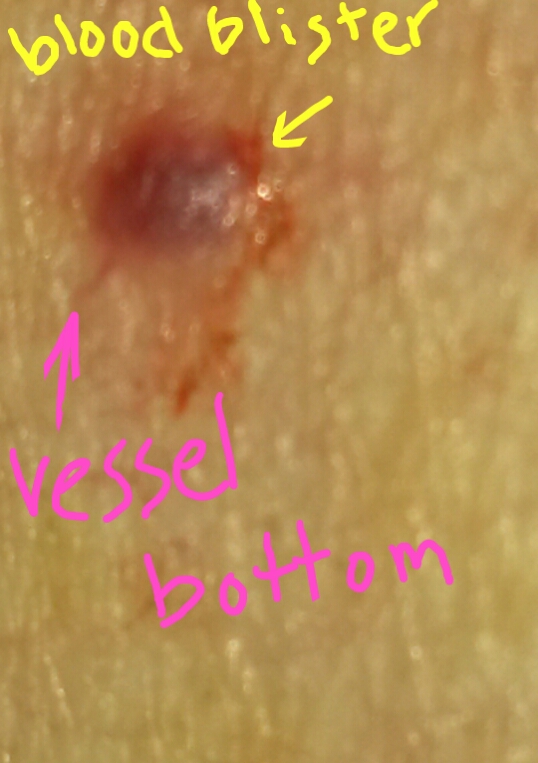
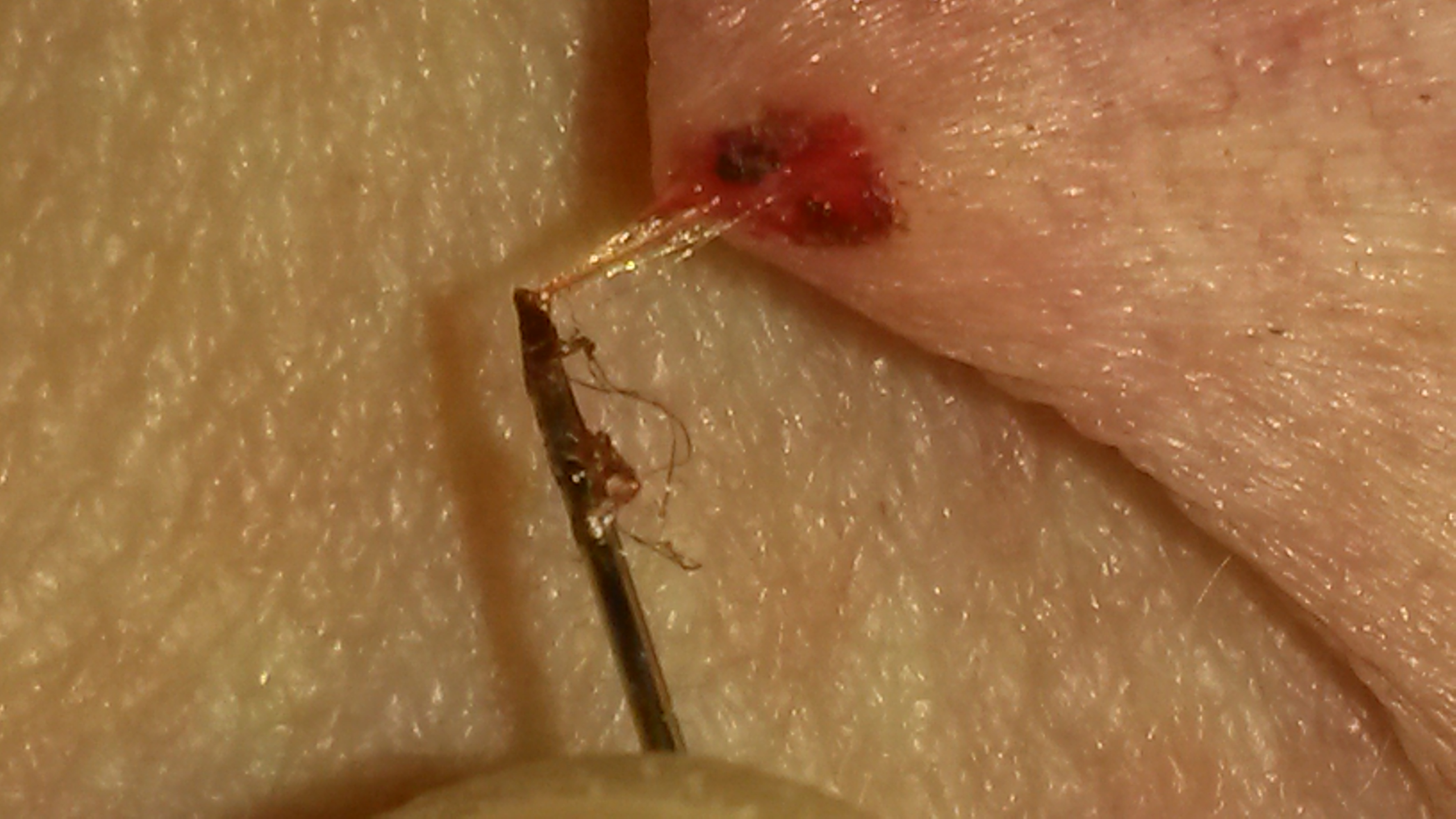
Below are a few photos showing a little more about The ©Pin Prick Shunt and how it works. In this photo here, it shows a blood vessel that our Founder knew was the location of the pressure build up in her upper left chest area. This is about 2" above her heart. The ©Pin Prick Shunt was placed less than a tap at first, then to 1/16mm inside where the arrow is pointing, this was a perfect location for entrance. You can see though where a blood blister that w as immediately formed, showed through the skin.
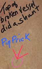
In these photos below, you'll see Photo #4 showing the formation with a yellow arrow pointing to the blood blister. -Tap Photo to see video---->
Looking at Photo #3 the blood vessel that looked broken from the top, was actually a solid Fibrelos Rock Clot that was a Vessel Clog all along. Once the blood blister formed, a few taps would blow out Fibrelos Rock Clots, you will see this in the video.
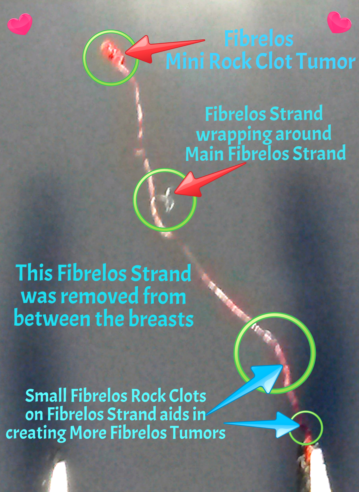
Photo #1 shows the ©Pin Prick Shunt in motion as Fibrelos Strands are being removed.
In Photo #6, a Fibrelos Strand is diagrammed.
In Photo #7, the ©Pin Prick Shunt is lifting hundreds if not thousands of Fibrelos Strands from Fibrelos Tangles, beneath the surface. Without this special technique using the ©Pin Prick Shunt for this removal, it's possible in would have stayed there forever.
Doctors, we are making you all aware of this new technique to further assist your Patients. Furthermore, once the prototype of the first ©Pin Prick Shunt is made, well note it on this website.
 White Flower Productions Copyright for this site: 2000 - 2022
All media is protected under this ©Copyright
©2000 - 2022 Copyright "©The Branchworm Study - Phase I" , "©The Fibrelos Study - Phase II" & "©The Fibrelos Dry Study", "©Fibrelos" is under Copyright ©2000 - 2022 for the ©Fibromyalgia Lyme Disease Foundation (©F.L.D.F.), "©The Pin Prick Shunt" ©2000 - 2022 is under Copyright for the F.L.D.F. ", ©The Natural Flowing Biopsy", ©2000 - 2022 is under Copyright for the F.L.D.F. , ©2018-2022 The Lyme-No-Mo & Lyme No More Campaign,©2020-2022, Copyright Fibrelos Airborne Particle Research Abstract ©2020-2022